New Electron Microscopy Tech Breaks into the Elusive Realm of Small Molecules
Researchers developed a laser-based phase plate for cryo-electron microscopy, enabling sharper images of small molecules like hemoglobin, previously difficult to capture with existing systems.
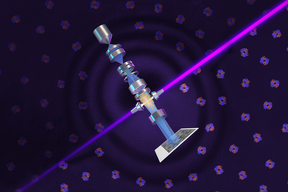
Nearly a century after the phase-contrast microscope revolutionized light microscopy, a team from UC Berkeley and Lawrence Berkeley National Laboratory has adapted the technique for cryo-electron microscopy (cryo-EM), which offers 10,000 times greater magnification. Their laser-based phase plate, detailed in a Science paper, produces clearer images of molecules that standard cryo-EM systems struggle to resolve, addressing a long-standing challenge in structural biology.
The system, named Theia, combines the phase plate with a custom Thermo Fisher Scientific microscope designed to maximize its laser’s ultra-bright output. Initial tests on proteins like aldolase and hemoglobin demonstrated improved resolution, particularly for smaller molecules. The technology also simplifies sample preparation by reducing interference from ice contamination, easing a time-consuming step in single-particle analysis that requires freezing samples to -160°C or below.
Researchers are now expanding Theia’s applications to cryo-electron tomography (cryo-ET), which reconstructs 3D images from multiple angles, akin to CT scans. This method captures molecules in their natural cellular context, offering higher resolution than light microscopy and addressing limitations of traditional single-particle cryo-EM. The team aims to enhance the prototype’s focus, potentially doubling structural information per image.
The project, supported by NIH, NSF, and other funders, reflects 15 years of collaboration among physicists, machinists, and biologists. The phase plate’s development involved trapping a 75-kilowatt laser in a mirrored cavity to achieve the precise phase shift needed for electron beams. While the current system requires specialized training, future iterations aim to streamline the technology for broader use in structural biology research.